



Restoring Dysfunctional Brain Circuits in Depression

By Conor Liston, M.D., Ph.D.
Robert Michels M.D. Professor of Psychiatry
Weill Cornell Medical College
BBRF Scientific Council Member
2013 BBRF Young Investigator
I'm a neuroscientist and a psychiatrist, and much of the work in my group has been oriented around answering an important unanswered question in depression neuroscience: What are the mechanisms that control the changes in mood over time that are the defining feature of depression?
Depression, by definition, is fundamentally episodic: it is defined by periods of low mood interposed between periods of wellness.
To date, most research has tended to focus on comparing groups of depressed people with groups of never-depressed people—i.e., “healthy controls”—and doing so at a single point in time.
We've learned a lot from this work. But it remains unclear why a person gets depressed today as opposed to last week or next month, and what determines how long they stay depressed. It’s also unclear, when depressed patients are treated and get better, what mechanisms initiate that recovery. We also don’t yet understand whether there may be other mechanisms involved in sustaining that recovery over time.
If we can advance our understanding of these questions, we can perhaps develop better ways of treating people and achieving more durable recoveries for our patients. The research I’m going to discuss here pertains directly to these questions.
HOW NEURAL CONNECTIONS ARE MODIFIED BY KETAMINE TREATMENT
My lab has been following people with depression and repeatedly getting functional MRI (fMRI) brain scans from them as they cycle in and out of depressed mood states, and then doing analogous studies in mice, whose brains we are able to study closely with sophisticated microscopes—something we obviously can’t do in people living with depression. The imaging technique we use for mouse brains allows us to look at how microcircuits in the brain change in response to chronic stress and in response to antidepressants. It’s likely that many mechanisms are involved in mood changes, but we (and others) think an important one involves the remodeling of tiny structures called postsynaptic dendritic spines. We often refer to them by the shorthand, “spines.”
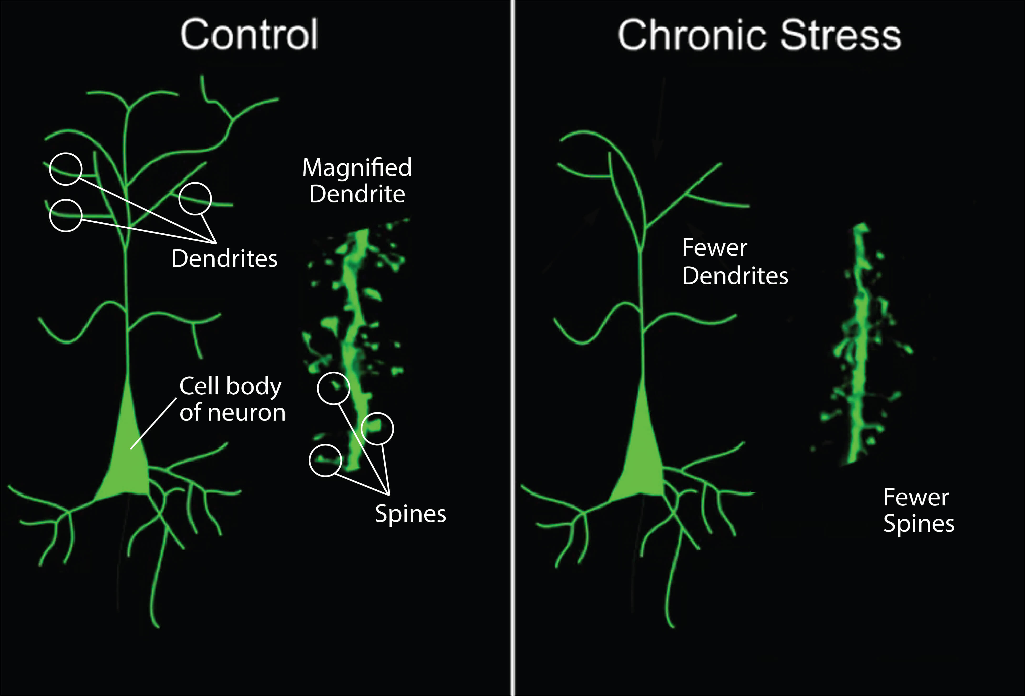
As you can see in the image above, spines are thorny protrusions from the branches (called dendrites) of neurons. You can think of each spine as a structural marker of a synapse—that is, a point of connection between brain cells.
We know that chronic stress, which can trigger depressive episodes in vulnerable people, reduces the number of these spines and therefore the number of of brain connections. Conversely, we know that drugs like ketamine, which act as antidepressants, do the opposite. They lead to the formation of new connections between brain cells.
Knowing this, it’s been tempting to speculate that the formation of new connections is required for initiating the mood improvement that many paitents experience when treated with antidepressants. Proving this would be an important advance. What is the evidence? In a first series of studies I'm going to describe, we set out to test this hypothesis.
Are the new connections we see in the depressed mouse brain after treatment with ketamine required to drive changes in mood and changes in behavior that we see shortly after the animals are treated? We found that they were required, but not in the way we expected. I'm going to try to explain what this means for depression treatment.
We use a technology called two-photon microscopy to observe the brain in great detail in living mice, including those exposed to stress, which can trigger depressive episodes in vulnerable people. This enables us to visualize in real-time the remodeling of dendritic spines, these points of connection between brain cells. The technology is amazing: we can return again and again to the exact same position in the brain with micrometer precision (millionths of a meter). And then we can ask what impact stress and antidepressants have on the remodeling of these brain cell connections.
One experiment involved imaging the brain before and after various chronic stressors. We asked what impact these stressors have on remodeling brain cell connections.
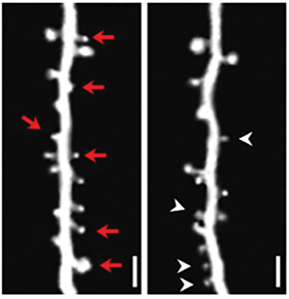
You don't need to be an expert to see the result—you can appreciate it by eye. In the pair of images above, the left image is a view of spines before an animal was injected with stress hormone, which mimics the impact of stress. In the right image, the same dendritic segment in the same animal 10 days later.
The red arrows in the left image above denote spines that were eliminated after the animal was stressed. You see them in that image, but not in the right one, after the equivalent of chronic stress. Conversely, the white arrowheads in the right image point to newly formed spines that weren’t present in the pre-stress (left) image.
We found that stress enhanced the rate of both spine elimination and new spine formation in multiple cortical areas. (New spines are routinely being created, especially early in the life of a mouse—or human being—as the brain is developing). But importantly, we found that chronic exposure to stress led to a net loss of spines.
It was interesting to us that the loss of connections didn't occur randomly. They occurred in specific positions in the neuron, which tend to cluster together in particular locations. This suggested to us that specific connections to the cortical cells we were looking at—specific projections from other brain regions—might be targeted for pruning by chronic stress, in a guided way. This led us to wonder if perhaps antidepressants were doing the opposite.
We treated the same group of stressed animals with ketamine or a placebo and then imaged them a day later. We chose that time point because, as anyone who has experience with ketamine treatment knows, after a single day, many patients report feeling that their mood is much improved after only one treatment. We found that in the ketamine-treated animals, there was a significant growth of synapses, a growth of spines, a net increase in connections between brain cells, that occurs after treatment.
This is consistent with the idea that ketamine (and possibly other antidepressants) might be acting to reverse the effects of stress. But this is something that needs to be tested.
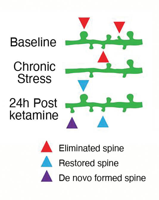
As shown in the schematic above, we classified each connection that formed after ketamine treatment. A “restored spine” [blue arrowheads] was one that formed in a position where a spine used to exist, pre-stress, and then disappeared with chronic stress. We also labeled brand new spines, which we call “de novo spines” [purple arrowhead]. These are spines that formed after ketamine treatment in a position where we didn't observe a spine before.
Ketmaine is restoring many connections that were lost after stress: We noted that about half of the connections that formed after ketamine treatment were restored spines. But at the same time, about half of the new connections we saw post-treatment were forming in new, random positions. This enables us to estimate that ketamine is partially rescuing the loss of connections that occurs after stress, but not fully rescuing it.
We think this might be relevant for patients' clinical experiences in a way that I'm going to describe next. Please keep in mind that one of the major challenges associated with ketamine is sustaining recovery from depression after the initial treatment.
AN UNEXPECTED FINDING ABOUT KETAMINE’S IMPACT ON CONNECTIONS
What we learned about the role of new connections that form after ketamine treatment was really interesting and not what we expected. We did two experiments. First, we imaged the mice before and after chronic stress, and then again in the hours and days, up to a week, after ketamine treatment. We asked which comes first: changes in the spines and their connections, or changes in the animals’ behavior?
We found that the changes in behavior—ketamine’s rapid antidepressant effect—occurs very quickly, just three hours after treatment. In fact, we now know that they occur even faster than that.
Whereas the formation of new connections didn’t begin until 12 hours after ketamine treatment, and peaked about a day later. This means that the formation of these new connections couldn't be required for initiating ketamine's antidepressant behavioral effects—because the improvement in behavior comes before the new connections form.
Another bit of data helped us understand more. It told us that the restoration of lost connections was tightly correlated with the maintenance of ketamine's antidepressant-like behavioral effect when we tested it two days or one week later. This enabled us to hypothesize that the new connections after ketamine are required for sustaining the antidepressant effects over time, even if they are not required for intiating these effects.
To test this, we teamed up with a group at the University of Tokyo that developed a technology that can be used to delete newly formed connections in the brains of animals in a very selective way. We used it to delete the newly restored connections after ketamine treatment and asked what impact it had on behavior. We found that if you delete those new connections, ketamine’s antidepressant behavioral effect wears off within a day or two.
We conclude from this and related experiments that after treatment with ketamine, the formation of new connections in this brain circuit isn't required for initiating an antidepressant effect, but it is required for sustaining it over a period of days.
This is important because we know that if you treat depressed patients with ketamine, many or most of them will report feeling a lot better the next day. But if you don't do anything else to help them and you just wait a week or two, almost all of those people will get depressed again. The impact that the new connections have, post-treatment, in sustaining the antidepressant effect wears off. That's why in clinical practice, ketamine is often delivered multiple times.
CAN WE EXTEND KETAMINE’S ANTIDEPRESSANT IMPACT?
Other research has shown that repeated ketamine dosing can be beneficial for boosting the formation of new connections, compared with a single treatment. In a clinic, ketamine may be administered twice a week for a few weeks. We know that with repeated dosing a larger proportion of those new connections will get integrated into circuits and, at least in mice, they can persist for the lifetime of the organism.
There might be other ways of maintaining the new connections that are forged after an initial ketamine treatment—ways that don’t involve repeated ketamine dosing. Exercise is known to boost the formation of new connections in the brain, and can assist with learning and memory. You can imagine that pairing exercise, at least in some individuals, with ketamine treatments (not at the same time, but perhaps in sequence on different days during the course of treatment) could be useful. We have preliminary data in animals suggesting this might be the case and it requires further study.
Pairing ketamine with psychotherapy is another possibility. The formation of new connections after ketamine treatment might render the brain more plastic, more responsive to the learning that takes place during psychotherapy, including the learning of new skills for promoting stress resilience. In treatment-resistant patients, we might further consider pairing ketamine with other treatments that spur connections, like TMS (transcranial magnetic stimulation), which stimulates the brain non-invasively through the scalp using magnetic-field pulses. It will be interesting to test if this gives rise to a synergy—benefits that one doesn’t get with either treatment given by itself.
DO OTHER ANTIDEPRESSANT TREATMENTS SPUR NEW CONNECTIONS?
Do other antidepressant treatments work as ketamine does to promote formation of new connections? We have evidence that at least some do. For example, new research from our lab shows that TMS treatments in mouse models of depression also spur new connections. TMS does so in a very specific subtype of cell, which is interesting. There's a lot of interest in psilocybin and other psychedelic compounds. Preliminary clinical trial data looks very promising. More research on psilocybin needs to be done, but there’s evidence that psilocybin also promotes the formation of new connections.
What we don't know yet is whether first-line, slower-acting antidepressant drugs like the SSRI medicines induce new connections in the way I’ve described here in the case of ketamine. Some researchers think this may be the case, but suspect they do so via different mechanisms and on a slower time course. Overall, I think there is good reason to think that new connections are a common pathway that is important across a range of antidepressants for maintaining antidepressant effects.
You might be wondering: What initatesthe antidepressant behavioral effects of ketmaine—the lifting of depression symptoms, sometimes within minutes? This has long eluded us. In recent months, our team has published results of experiments that we think demonstrates the mechanism behind the drug’s rapid action. This research has potentially important implications for improving the treatment of depression, including the development of new rapid-acting antidepressants with fewer side effects than ketmaine. [Editor: full details of this important new research will be the subject of a story in our next issue.]
HOW NEURAL CIRCUIT FUNCTION IS MODIFIED BY KETAMINE
I'd like to talk a bit about experiments we did in parallel with the research on new connections I’ve already discussed, focusing on what we see in terms of circuit function in animals undergoing antidepressant treatments. To test this, we use a method very similar to the one I have described: live, 2-photon imaging of mouse brain circuits through a cortical window. But instead of imaging the structural markers of connections between brain cells, the dendritic spines, in these experiments we're imaging the cells’ functional properties—not how they’re connected but rather what they are doing.
In the series of images below, which are actually individual frames of a movie we made, cortical neurons have been genetically engineered to glow green when they become active. In the movie, you get these beautiful images that look like Van Gogh's “Starry Night,” where neurons are lighting up and then disappearing and lighting up again. We try to suggest this in the row of individual frames from the movie, below.
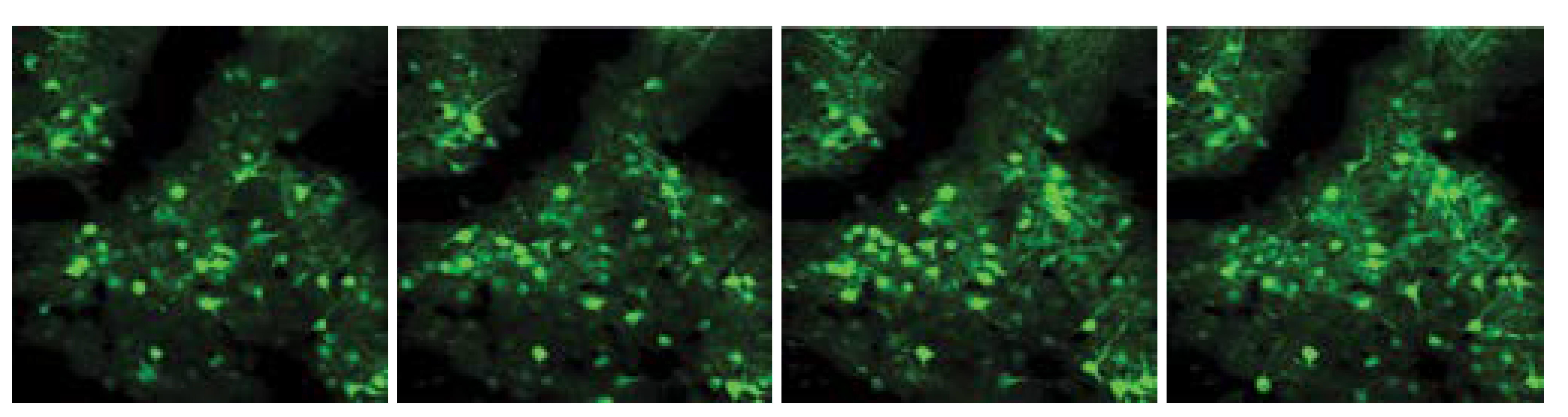
This visual readout serves as a marker of how active the brain cells are. We predicted that if many connections between neurons were being lost after chronic stress and restored after ketamine treatment, then we might see changes in what we call functional connectivity—the degree to which the activity in connected pairs of cells is correlated. We would expect to see activity levels change after chronic stress and then go back to normal after ketamine treatment. And that's indeed what we did observe.
Using color-coding, we were able to see and compare activity in cells that are strongly connected functionally, and in cells that are not strongly connected functionally.
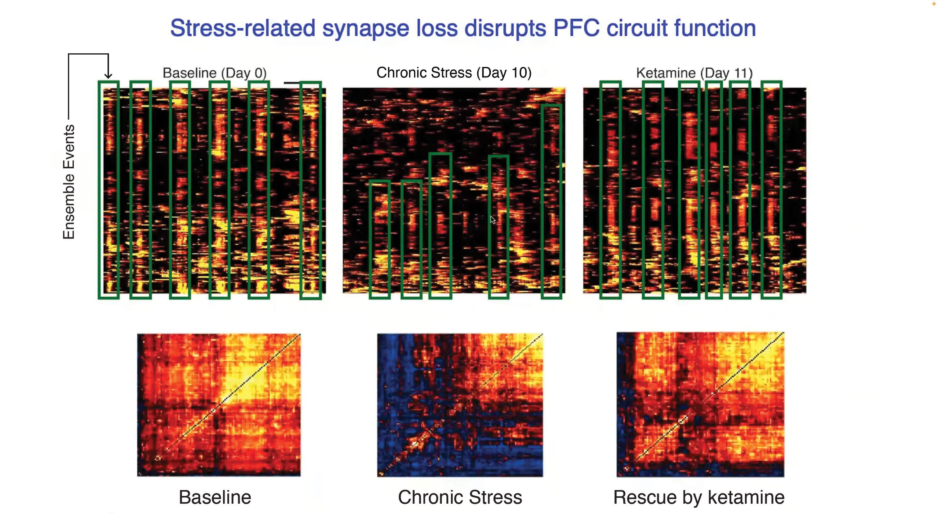
In the “heat-map” images above, warm colors denote pairs of cells that are strongly functionally connected, and cool colors denote pairs of cells that don’t have a strong functional connection. The baseline, prior to stress, is seen in the lower left panel. In the lower middle panel, after chronic stress in this animal, there's a loss of warm colors (lower middle panel), which means a loss of functional coupling between neurons. When we treat the animals with ketamine, those warm colors come back (lower right panel).
The reason we see these changes has to do with what we call “ensemble events.” These are visualaized in the 3 corresponding images on top, in which you read the data in columns, from top to bottom. A lot of the activity we captured occurs when these ensembles of cells become active at about the same time. And you can see that chronic stress (top center panel) significantly disrupts the occurrence of these ensemble events (and when they do occur, they tend to involve fewer cells). In the top right image, you can see that after ketamine treatment, the ensemble activity we saw before stress is restored.
We think these ensemble events are behaviorally relevant in various ways. We think they impact fear learning, cognitive flexibility, the perception of social hierarchies, and reinforcing social interaction behaviors. But next, I'm going to focus on another behavioral impact, pertaining to neural activity related to seeking rewards, which is highly relevant in depression.
REWARD CIRCUITS, ANHEDONIA, AND KETMAINE
How do you respond when you're motivated to obtain a reward? You make an effort to obtain it. We know from past experiments that a particular circuit in the brain, which projects from the anterior cingulate cortex (ACC) to an area involved in reward processing called the nucleus accumbens (NAc), is important for encoding a predictive signal of reward—our expectation of receiving a reward, given various levels of effort. The ACC—>NAc circuit integrates information about effort to gain a reward. Its signal is required for reinforcing future decisions to expend effort to obtain rewards.
The signal in this circuit scales with effort. We think this is really relevant in depression, because we know that patients often experience anhedonia, a loss of interest or pleasure in things or activities that they used to enjoy.
You can imagine if you're depressed that even getting out of bed, getting dressed, walking to your favorite restaurant to spend time with friends, these things might feel very effortful and you might just choose never to engage in them. But we also know that if you encourage a depressed person to actually engage in the enjoyable activity, they often do enjoy it—the problem often lies in willingness to expend effort to obtain the reward.
We think that the ACC—>NAc circuit I mentioned is very important for those kinds of decisions. It turns out that in animal experiments, after chronic stress, the animals very consistently stop choosing high reward/high effort options in favor of a low reward/lower effort option.
We're currently looking at how antidepressants like ketamine influence the function of this circuit.
DO NEURAL NETWORKS CHANGE AS PATIENTS’ DEPRESSION SYMPTOMS CHANGE?
Next, I’d like to tell you about our work in people with depression. We're very grateful to them for making this work possible. Very dedicated reseachers in my lab tracked a small number of people 40, 60, up to 120 times, over a period of 1–2 years as the patients cycled in and out of depression.
They collected brain scans from these participants over and over again, along with detailed clinical assessments that allowed us to map what is changing in the brain as a person's symptoms change.
You can see in the illustration below—again, data rendered in the form of a “heat map”—that there are a number of symptoms displayed in rows; they’re related to anhedonia, loss of interest in pursuing pleasurable activities. The darker colors depict times when this particular person was more anhedonic. The lighter colors denote times when this person didn't really have much anhedonia at all.
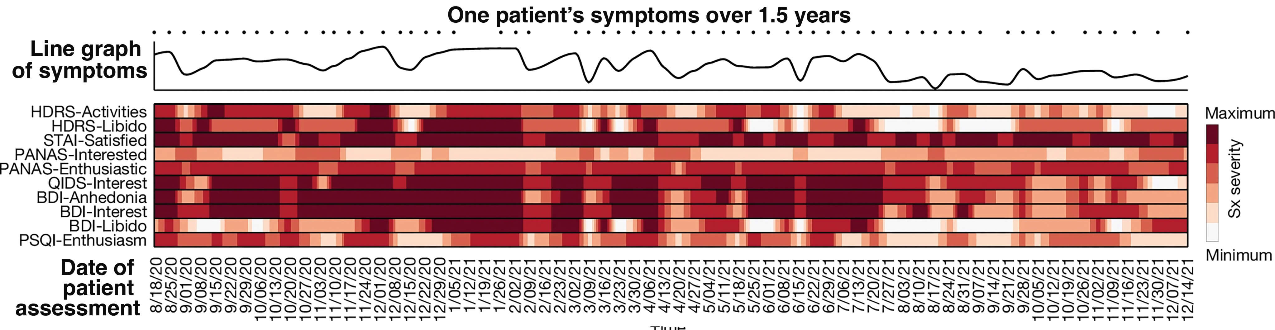
What you can see by glancing at the line graph just above the heat map, summarizing the data, is that there were a lot of fluctuations. Sometimes this person reported very severe anhedonia, but sometimes had essentially no anhedonia. Because we're doing this sampling repeatedly over time, we realized that we'd be able to study how these changes relate to changes in network topography—the boundaries of neural networks—as they change over time, as revealed in fMRI scans.
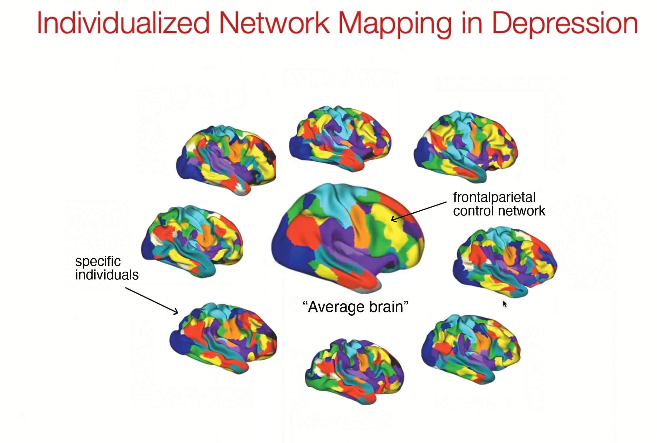
We've known for some time that the brain is organized into functional networks, which are depicted in the brains above by different colors. Recently, scientists have discovered that the boundaries of these networks are actually quite different in different individuals, kind of like a fingerprint or even like a face.
You can see that everyone whose brain is depicted in this graphic has a network we’ve colored yellow [see arrow] called the frontoparietal control network. Everyone has it in about the same position, but the boundaries are dramatically different across these different individuals. In some people, it's very small, in some people it's much larger.
Now think of this in the context of using an antidepressant treatment like TMS, which targets a particular area of the brain based on positioning a magnetic coil over a certain place on the scalp. In different patients this would potentially affect dramatically different networks, based on the network topography of their brains as shown in this graphic. We wanted to ask whether these networks are systematically different in the brains of depressed people compared to people who've never been depressed.
ENLARGEMENT OF THE SALIENCE NETWORK IN DEPRESSION
What we found was really striking: one network and only one network is dramatically expanded in the brains of people who suffer from depression. This network is called the frontostriatal salience network. The “salience network,” as we refer to it, is important in orienting attention to emotionally important or relevant stimuli in the environment.
We find that this network is dramatically expanded in depressed people. Again, you don't need fancy statistics, you can just see it. In the set of brain images below, the salience network (shaded in black) is much larger in three individuals with depression compared to a never-depressed person. We see this very consistently across many people with depression.
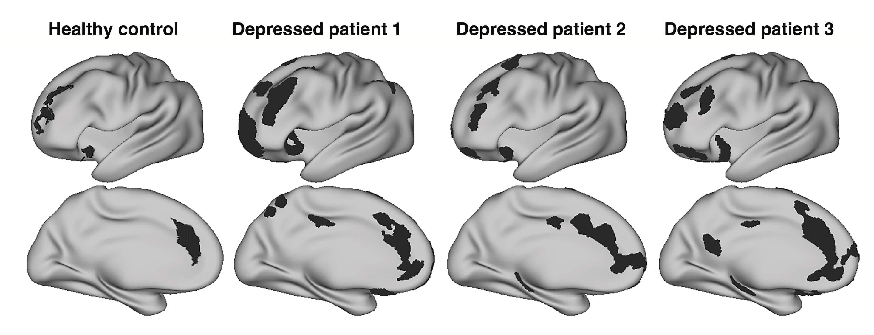
One question we asked was about how the expansion of the salience network affects orienting our attention to emotionally salient stimuli, and why this might be important—what it might do to contribute to depression.
Perhaps it's the case that this network expands as a person gets depressed and then contracts as a person gets well again. We were well positioned to test this hypothesis because as I’ve noted, we have imaging data as people became depressed and not depressed and depressed again. What this showed us was just the opposite of what we predicted.
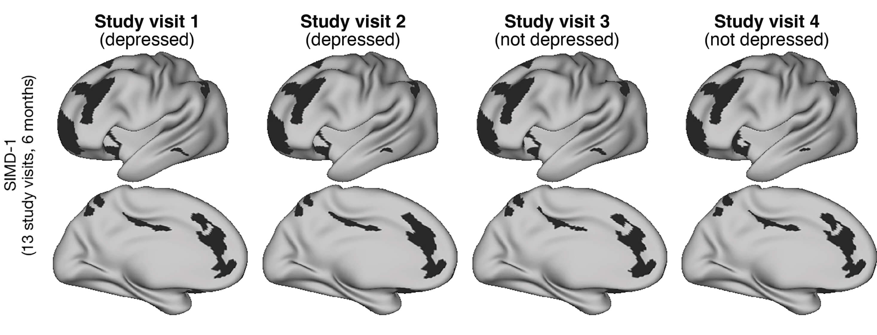
The graphic above shows the boundaries of the salience network (in black) in one individual, over four representative study visits. In the two early visits, shown on the left, the person is depressed. In two subsequent visits (right), the person is not depressed. You can see that the boundaries of this network hardly change over time. We see this in all the patients we scan: the network topography, the size of the salience network, really does not change. It's unrelated to how depressed they are currently at the moment. This led us to rethink our hypothesis.
We next had the idea that perhaps this salience network isn't expanding and contracting as a person gets depressed and not depressed, but instead that it somehow is conferring risk for depression.
How might we test this? We reasoned that if that were the case, we should find that the network is expanded in individuals who have never been depressed but later go on to become depressed. This, in comparison to the network’s size in individuals who were never depressed and don’t become depressed later on.
We turned to data in a unique resource called the ABCD study, which has been going on for years. Over 10,000 kids have been enrolled and then followed from ages as young as 7 up through their teen years. Among those 10,000 kids, we selected 60 who were never depressed at ages 10 and 12 and then went on to become depressed in their teenage years. Because of their participation in the ABCD study these children had brain scans at both of these time points.
We did indeed find that the salience network was expanded in the children who went on to become depressed a few years later—this, compared to the nework’s size over time in those who did not become depressed. We're currently investigating how an enlarged salience network might contribute to risk.
Importantly, we also found that the expansion of this network occurred differently in different individuals. When this network expands, it encroaches on the territory of its neighbors in different ways. You can cluster the people in the sample and identify at least three subgroups of patients based on which neighboring networks are being encroached upon.
We think that the increased risk conferred for depression may be related either to the expansion of the salience network or to how it encroaches on its neighbors, or some combination of the two.
We're currently incorporating our data into tools we've developed for subtyping patients with depression based on fMRI data. We already know that these fMRI derived subtypes are associated with different kinds of responses to TMS treatment for depression. This relates to what I mentioned earlier: people with different patterns of functional connectivity might be expected to respond differently to brain stimulation at a given site.
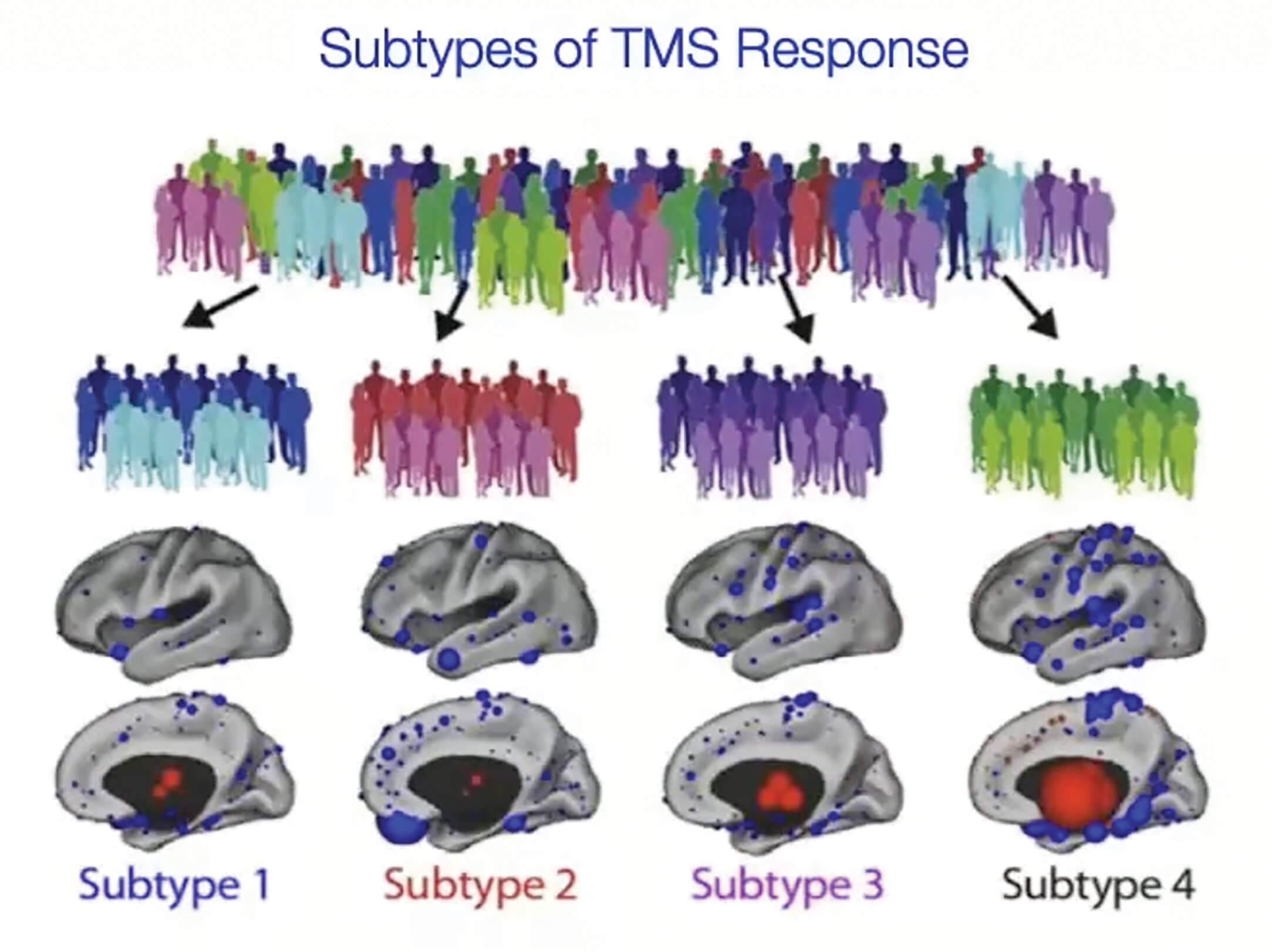
To give an example of how such information might be used, we found that one of the subtypes we identified, Subtype 1 (see graphic above) is highly responsive to TMS. Subtypes 2 and 4 are not at all responsive and subtype 3 shows an intermediate level of responsiveness. We now know that you can use a similar approach to model individual differences in response to SSRI antidepressant medciations.
We hope to be able to combine these findings into a pipeline that might be used, first, to identify people who are likely to respond to first-line SSRI medications for depression, and then, second, give them the medication that we think will work.
If we think that they're not very likely to respond to this medication, we could perhaps consider prioritizing second-line treatments like TMS and using this subtyping approach to identify what is the optimal target for treatment. We're currently testing this strategy to see exactly how well it works.
