



Accelerated TMS Guided by Individualized fMRI Connectivity Targeting Outperformed Conventional Targeting in Major Depression Patients
Accelerated TMS Guided by Individualized fMRI Connectivity Targeting Outperformed Conventional Targeting in Major Depression Patients
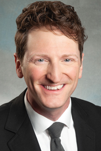

A clinical trial led by BBRF grantee and prizewinner Joseph J. Taylor M.D., Ph.D., of Harvard and Mass General Brigham, has generated head-to-head evidence that neuroimaging can be used to improve clinical outcomes in psychiatry. Prior research has suggested such efficacy, but not in patient trials directly comparing use vs. non-use of neuroimaging-based targeting to achieve therapeutic results.
The trial compared a neuroimaging-guided version of accelerated transcranial magnetic stimulation (aTMS) in treatment-resistant individuals with major depressive disorder with aTMS therapy targeted conventionally—based on measurements taken of the scalp.
TMS, pioneered by BBRF grantee and Scientific Council member Mark S. George, M.D., was first approved by the FDA in 2008 for treatment of depression. The technology is based on using magnetic stimulation delivered non-invasively by coils placed above the scalp to therapeutically modify the activity of neural circuits thought to be involved in depression. TMS has been modified and improved over the years. Among other innovations, it can be delivered on an accelerated schedule involving multiple stimulation sessions per day over the course of just one week, compared with single daily sessions over 4 to 6 weeks in the original TMS protocol.
The late Nolan R. Williams, M.D., a BBRF grantee and prizewinner, and a former student of Dr. George, spearheaded development of an accelerated and intensified TMS protocol called SAINT at Stanford University. In 2020, his team first reported that SAINT generated rapid antidepressant effects. In 2022, the FDA cleared commercialization of the protocol for patients with refractory depression.
In SAINT and a number of other accelerated TMS protocols developed in the early 2020s, neuroimaging is used in each patient to optimize the TMS target. Rather than measure the scalp as is done conventionally (finding a spot above the brain’s dorsolateral prefrontal cortex, or DLPFC), the SAINT protocol uses functional MRI scanning to find a spot in the brain beneath the scalp that, in functional terms, is most “anti-correlated” with activity in a deep-brain area called the subgenual anterior cingulate cortex (sgACC), implicated in depression. The target location varies somewhat from person to person.
Dr. Taylor, winner of BBRF’s Klerman Prize in 2025 and a 2022 BBRF Young Investigator, who is also a former student of Dr. George, and colleagues who included Shan H. Siddiqi, M.D., and Michael D. Fox, M.D., Ph.D., conducted a randomized, blinded clinical trial involving 40 individuals with moderate to severe major depression symptoms and treatment resistance. Dr. Siddiqi is the 2022 BBRF Klerman Prize winner and a 2019 BBRF Young Investigator.
The patients were in the 40s, on average, a little more than half were female, and 72% were White. The researchers compared conventionally targeted aTMS (i.e., targeted via standard scalp measurement) in half the participants with a modified version of aTMS in the other half that was targeted based on results of fMRI scans in each patient.
In lieu of the proprietary SAINT protocol, the team used a new targeting method that is also based on fMRI imaging. This method, which they have been developing for several years to potentially improve upon existing methods, takes advantage of public-source data on brain circuitry published by Drs. Siddiqi, Fox and colleagues in 2021. In that year, they found what they described as a common or convergent “casual circuit” in depression spanning many different brain regions. “This whole-brain circuit was derived from the connectivity profile of 14 independent datasets of brain lesions causing depression as well as TMS and deep-brain stimulation sites relieving depression,” the team explains.
Each patient receiving the fMRI-based aTMS treatment was targeted at the spot above the DLPFC that was most strongly connected to the previously identified convergent depression circuit. There is some retrospective evidence that such a target will outperform targeting the sgACC in patients with major depression, the team said, although there is no consensus on this point. Importantly, targeting the “convergent circuit” can be done for free based on publicly available data—potentially making it more scalable. One possible disadvantage is that, at least in this trial, each patient so targeted had a 41-minute-long multi-echo resting-state fMRI scan, which some potential practitioners are not equipped to give and some patients may not be able to afford.
The actual aTMS treatment given patients in both the scalp-measured and fMRI-scanned groups involved 10 brain stimulation sessions per day over 5 days consisting of 18,000 magnetic pulses per day (90,000 total). Live neuronavigation feedback was used in both groups, to keep the pulses on target.
Reporting results in JAMA Psychiatry, the team noted that at the primary endpoint of the study, 1 month after the conclusion of the aTMS sessions, therapeutic outcomes were superior in the group with connectivity-based targeting relative to that receiving scalp-based targeting. The antidepressant effect, as measured by the standardized MADRS symptom scale, was greater, and 80% vs. 60% achieved a clinical response (defined as a reduction in symptoms of 50% or greater).
In the team’s view, the most important result was that connectivity-based targeting “had a large effect size relative to scalp-based targeting.” Both, as noted, were clinically effective (80% vs. 60%), but in the group whose targeting was based on connectivity, the team calculated that one additional patient out of every 5 patients so targeted (vs. those conventionally targeted) had responded to the treatment at the study endpoint (30 days after the end of treatments). This particular measure of impact, said the team, “compares favorably” to measures of various diagnostic imaging techniques currently in use in other medical contexts.
The team concluded that their results justify replication in clinical trials involving larger patient groups. There were a number of caveats. Other trials may involve testing other neuroimaging-based targeting methods (e.g., PET and structural MRI). If so, “target reproducibility” such as they demonstrated within individuals will be important to replicate. “We found that our targets were reproducible within an individual and different across different individuals, [which are] both important if individualized targets are to be clinically useful.”
The 41-minute-long multi-echo fMRI scan in the current trial generates much more information than fMRI scans often used for targeting, and it was not clear based on this study whether so much data was really needed to achieve clinical advantages—something that must be further explored.
Perhaps most important, additional trials must confirm a clinical difference in antidepressant response, and might also explore whether different symptoms experienced in major depression may be best addressed using different TMS targets.
The team also included: Tracy Barbour, M.D., 2017 BBRF Young Investigator; and David Silbersweig, M.D., 1996 BBRF Young Investigator.
