PTSD Symptom Reductions in Clinical Trial Are Linked to Changes in White Matter
PTSD Symptom Reductions in Clinical Trial Are Linked to Changes in White Matter

A new analysis of the results of a clinical trial involving the treatment of people diagnosed with PTSD has yielded insights into how the tested treatment affected connectivity in the brain and how those changes, both structural and functional, in turn, correlated with reductions in PTSD symptoms. By providing a detailed picture of how a treatment worked upon the brain, the new findings are part of the larger effort to improve treatments and results for patients in the future.
Results of the new study, reported in the journal Psychiatry Research: Neuroimaging, extend analysis of a clinical trial led by Ilan Harpaz-Rotem, Ph.D., of the Yale University School of Medicine and the U.S. Department of Veteran Affairs National Center for PTSD and the VA Connecticut Healthcare System. Dr. Harpaz-Rotem, whose 2015 BBRF Independent Investigator grant helped support the trial, is senior author of the new paper; its first author is Nachshon Korem, Ph.D., also of Yale and the VA.
A total of 26 patients diagnosed with PTSD provided data included in the analysis of both the trial and this new study of its results. The average patient’s age was about 40, and 10 of the participants were female. Participants were given an infusion of either ketamine (an experimental drug for PTSD) or a control drug (midazolam). For four days following the scans and drug infusions, participants received 90- to 120-minute sessions of exposure therapy given by a clinician. Brain scans were made before treatments began and again following the treatment course on day 7, and again 30 days post-treatment.
The treatment they received, on average, reduced participants’ score on a standard measure of PTSD symptoms called the CAPS-5 scale by about 17 points, at the end of the 30-day post-treatment follow-up. The scale assesses individuals on the basis of 20 questions, each scored from 0 to 4, meaning the range of possible scores is 0-80. Trial participants’ mean pre-treatment CAPS-5 score was about 48 and their mean score at the 30-day post-treatment follow-up was about 31. A score of 31-33 is usually regarded the threshold for a PTSD diagnosis.
The new study followed upon results obtained in the clinical trial. It was designed to test a specific hypothesis: If brain activity underlies behavior, then sustained changes in behavior such as symptom improvement should reflect lasting changes in the brain. As pointed out by Dr. Korem, “functional activation patterns of neural circuits in the brain are inherently transient and sensitive to context, mood, and internal state. For these changes to support long-term behavioral change, they must be consolidated into more stable anatomical alterations.” The new study tested this hypothesis by examining both functional and anatomical connectivity in the brain, before and after treatment.
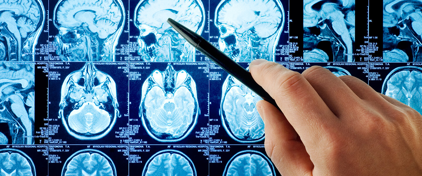
Each of the trial participants received brain scans that included structural MRI, diffusion tensor imaging (DTI), and functional MRI (fMRI). Much information about brain anatomy can be gleaned from MRI; the DTI scan reveals the structure of white matter in the brain—consisting mostly of the fatty material called myelin that insulates the axons which connect neurons. In combination, analysis of the scans made in the trial focused on functional and anatomical connectivity within the same white matter tract, the uncinate fasciculus (UNC), before and after treatment. They sought to understand the relation between structural and functional circuit connectivity as revealed by the scans, and corresponding changes in PTSD symptom severity as modified by the combined drug and exposure therapy treatment.
In their paper the team says that “for the first time, we [were able to] show how short-term changes in functional connectivity are associated with short- and long-term changes” in a measure of white matter integrity called FA (fractional anisotropy) in the UNC, which is implicated in giving rise to PTSD symptoms.
Specifically, the team discovered that reduced white matter integrity in the UNC was associated with a reduction in PTSD symptom severity. The reduction in white matter integrity in the UNC after treatment was specifically linked with a decrease in connectivity between the brain’s hippocampus and the ventromedial portion of the prefrontal cortex (vmPFC).
This finding, the team said, supports what they term a “network neuroscience approach to understanding what goes wrong in the brain in PTSD and how dysfunction can be reduced with successful treatment.”
The network neuroscience approach, the team notes, posits that “recurring changes in functional connectivity induce changes in white matter, which in turn facilitate lasting behavioral transformation.” The alterations in functional-anatomical connectivity may be driven by the recurrent exposures [to the traumatic memory] enacted during therapy. When therapy alters hippocampus-vmPFC connections, they have the effect, the team hypothesizes, of therapeutically altering emotional processing and emotional regulation, as well as regulation of the process by which fear memories are retrieved by an individual.
The team called for more research along the same lines, in part to address the two main limitations of their study of the clinical trial data: one, the relatively small number of patients in the trial, and two, the fact that the treatment being assessed involved the administration of two drugs (ketamine and midazolam) in addition to extended exposure therapy. The impact of the drugs upon the symptom reductions and connectivity changes that were observed, if any, was impossible to deduce based on the trial data.


